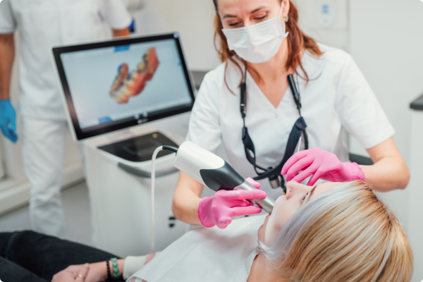
Intraoral scanners offer an accurate and fast digital way to take dental impressions. They are the ideal starting point for every dental treatment which requires an accurate and detailed representation of the surface of patients’ teeth and gums.
Introduction
The technology behind intraoral scanners has evolved to make them a vital piece of equipment that improves diagnosis and treatment planning as well as increasing business efficiency and patient satisfaction.
The popularity of this technology is reflected in the fact that the global intraoral scanners market continues to grow, being valued at $355.8 million in 2020 and estimated to reach $704 million in 2027[1].Is it finally time to put down your impression tray?
What is intraoral scanning?
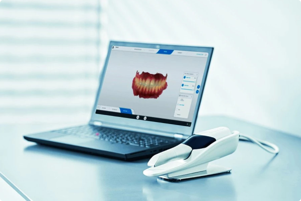
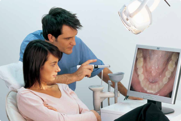
Intraoral scanners (IOS) are devices for capturing direct optical impressions using light beams or lasers projected onto the surfaces in the mouth. A small camera fitted to the end of a handpiece is passed over the teeth, gingiva and arches and the digital software immediately constructs a 3D digital model of the subject on screen.
The predictability and accuracy of a digital workflow virtually eliminates the need for remakes, makes considerable savings on lab fees and does away with the additional costs of impression materials. There is also a considerable environmental impact in that we are no longer using numerous plastic trays and large amounts of silicone, all of which has to be disposed of

The predictability and accuracy of a digital workflow virtually eliminates the need for remakes, makes considerable savings on lab fees and does away with the additional costs of impression materials. There is also a considerable environmental impact in that we are no longer using numerous plastic trays and large amounts of silicone, all of which has to be disposed of
Single-visit restorations
A common application for intraoral scanners is the first step in a chairside same-day restoration, such as a crown or veneers. In-practice CAD software has become increasingly sophisticated, as well as easy to use, so that, once the intraoral impression has been imported, it can provide viable computer-generated restorative proposals, which the clinician can adapt and refine. This makes designing restorations remarkably straightforward and enables the clinician to keep more control of the design process.For simple restorations such as veneers, inlays, onlays and single crowns, it is then possible to mill the restorations on an in-practice milling unit. A chairside setup like this gives a clinician significant CADCAM capabilities and ultimate flexibility which allows them to control whether they keep the full process in-house or pass more complex cases on to the lab.
Practice-lab workflow
For more complicated restorations, or if chairside restorations are not an option, clinicians can design their restorations and send over those designs to the lab. Alternatively, they may choose to take the digital impression and send the files straight over to their preferred laboratory for both the design and manufacturing stages.Most intraoral scanners can export scan images in an open STL format which means they can be imported into software from other manufacturers, giving clinicians the flexibility to choose the laboratory that best suits their requirements.
Most CADCAM software also integrates with related software that enables clinicians to design orthodontic appliances or send digital impression files straight to third party orthodontic providers such as Reveal® and Invisalign®. With the addition of a 3D printer to the practice, items such as orthodontic aligners and surgical guides can be manufactured in the practice as well.
How to evaluate intraoral scanners
There are many factors to consider before investing in an intraoral scanner. All of the following are recommended criteria:
- Open and therefore able to work with a variety of systems
- Detailed, full colour, 3D or higher visual representations
- Regular software updates
- Clinically proven to be accurate
- Fast
- Comfortable for patient and operator
- Cost-effective
- Long warranty and good customer service
- Future-proof / easily upgradeable
- Easy to use and keep clean
- Powderless
What intraoral scanners are available with HS?
;)
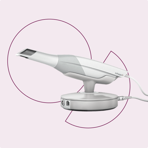
3Shape TRIOS 5
Combine technology, ergonomics, ease-of-use, and hygiene
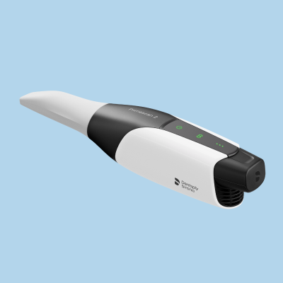;)
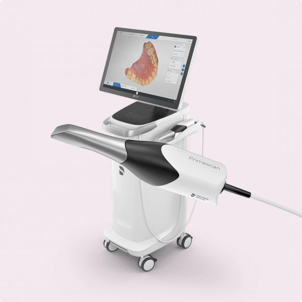
Primescan 2
The new wireless intraoral scanner is simple and versatile, easily integrating into your practice's existing setup to help you grow your practice and deliver exceptional patient care.
The Power of Digital Scanning and X-ray Imaging Combined
Digital impressions and digital X-rays both deliver extraordinary accuracy and clarity and have revolutionised impression and X-ray taking respectively. But what happens when the two are combined?
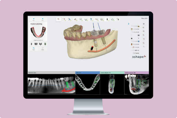
CADCAM software can merge the surface scan from a digital impression with a 3D digital X-ray to give the clinician a complete picture of the oral situation above and below the gum line. This is invaluable for some areas of dentistry in particular and also for communication both with the patient and within the dental team.
Implant-borne restorations
Combining a surface impression from an intraoral scanner with a 3D digital scan gives an implant surgeon all the information required to plan the placement of one or more implants and restorations. The impression and digital scan are imported into CADCAM software and merged. The clinician designs the restoration within the software and plans the implant placement concurrently, ensuring they work seamlessly together. They can even design a surgical guide within the software and print it out on a 3D printer, or order it along with the restoration from the laboratory. This means that implants can be placed and the prosthesis manufactured with pin-point accuracy which results in an accurate, aesthetic and long-lasting restoration.Orthodontics
Digital impressions and 3D digital X-rays can be used together to deliver custom, patient-specific orthodontic appliances – even in-house with the addition of a 3D printer. A 3D radiographic view of the patient’s anatomy means the clinician no longer has to guess the 3D location of an impacted tooth or the correct eruption vector. Merging the X-ray and the surface scan enables the clinician to use the root anatomy to devise the correct orthodontic treatment plan, improving the long-term outcome.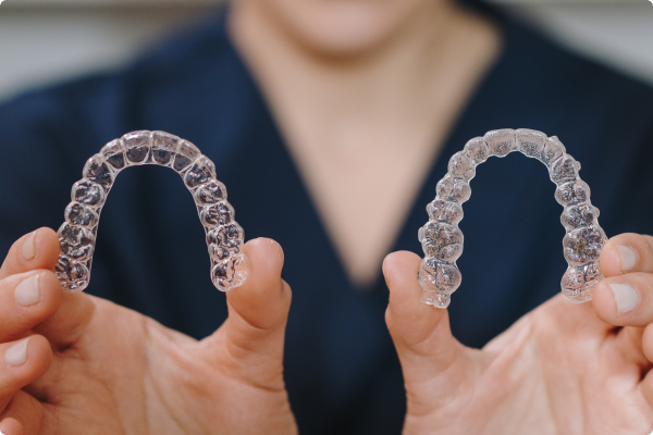
Patient communication
The ability to show the patient images of their oral anatomy both above and below the gums gives patients a much better understanding of the diagnosis and the intended treatment plan. Accurate pictures speak more than a thousand words which significantly improves patient understanding and therefore treatment uptake.Dental team communication
Digital images and CADCAM software have succeeded in improving communication between the whole dental team, including clinician, technician and any third-party partners. Communication is instant and misunderstandings are avoided because every team member has immediate access to the detailed images and design files. From a patient record-keeping point of view, digital images make it much easier to keep all patient information together in a single file on a practice management system for instance. Diagnostic images can then also be tracked more easily to monitor disease progression or any other changes in the patient’s dentition.