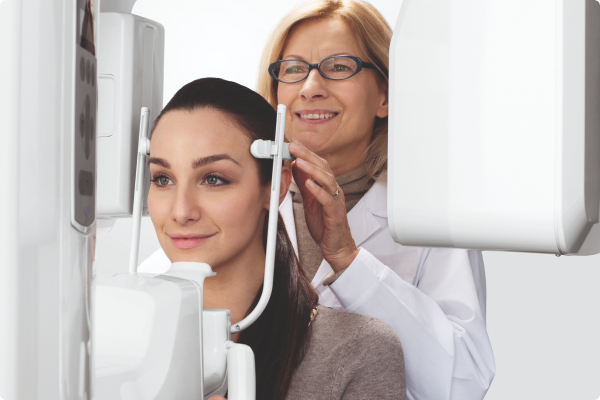
What is digital X-ray imaging?
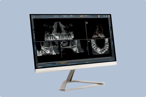
X-ray imaging is the taking of X-rays of the mouth to generate images of the tooth and bone structure, soft tissue and nerve paths underneath the gums. Digital X-rays are taken by machines which have a digital image sensor which captures the X-ray and displays them on a digital screen. They are fast replacing traditional film X-rays due to the lower doses of radiation required and the impressive level of detail which can be achieved with digital technology.
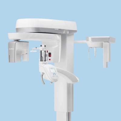
There are two types of digital X-rays commonly used in dentistry, 2D digital X-rays and 3D X-rays. Traditionally in an effort to keep radiation doses as low as reasonably achievable and because 3D X-rays have until now required much larger doses of radiation, 2D digital X-rays have been widely used.
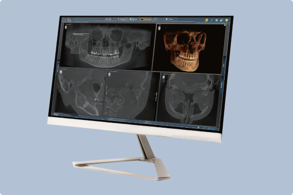
However, the technology behind Cone Beam Computed Tomography (CBCT) scanners, which are used to take 3D digital X-rays, has improved markedly recently and it is now possible to take extremely clear and detailed images at very low doses of radiation.
Dental practices are required to adhere to both the Ionising Radiation (Medical Exposure) 2000 Regulations (IR(ME)R) and the Ionising Radiations Regulations 2017 (IRR17) , which are based on the ALARA principle of only exposing a patient to a radiation dose that is “as low as reasonably achievable.” As a rule, dental practitioners should favour a lower dose wherever practicable, as long as the resultant X-ray provides the detail and information required to make an accurate diagnosis and an effective treatment plan.
How to evaluate digital X-ray machines
There are many factors to consider before investing in a digital X-ray machine. All of the following are recommended criteria:
- Open and therefore able to be used by a variety of dental equipment and software
- Clinically proven
- Cost-effective
- No ongoing fees or licences
- Long warranty
- Future-proof / easily upgradeable
- Offers a range of definition settings and low dose options to cover all applications
- Offers a range of fields of view to ensure adherence to the ALARA principle
- Easy to use, including straightforward patient positioning
Browse Our Best-Selling Digital X-ray Machines
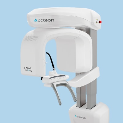;)
Acteon X-MIND Prime 2
Get a complete set of panoramic exams fast for optimal patient diagnosis
Acteon X-MIND Prime 2
Get a complete set of panoramic exams fast for optimal patient diagnosis
;)
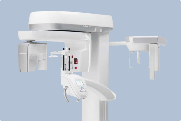
NewTom GiANO 3D
Wide choice of examination options enable a broad scope for diagnostics
The Power of Digital Scanning and X-ray Imaging Combined
Digital impressions and digital X-rays both deliver extraordinary accuracy and clarity and have revolutionised impression and X-ray taking respectively. But what happens when the two are combined?
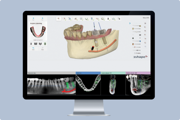
CADCAM software can merge the surface scan from a digital impression with a 3D digital X-ray to give the clinician a complete picture of the oral situation above and below the gum line. This is invaluable for some areas of dentistry in particular and also for communication both with the patient and within the dental team.
Implant-borne restorations
Combining a surface impression from an intraoral scanner with a 3D digital scan gives an implant surgeon all the information required to plan the placement of one or more implants and restorations. The impression and digital scan are imported into CADCAM software and merged. The clinician designs the restoration within the software and plans the implant placement concurrently, ensuring they work seamlessly together. They can even design a surgical guide within the software and print it out on a 3D printer, or order it along with the restoration from the laboratory. This means that implants can be placed and the prosthesis manufactured with pin-point accuracy which results in an accurate, aesthetic and long-lasting restoration.Orthodontics
Digital impressions and 3D digital X-rays can be used together to deliver custom, patient-specific orthodontic appliances – even in-house with the addition of a 3D printer. A 3D radiographic view of the patient’s anatomy means the clinician no longer has to guess the 3D location of an impacted tooth or the correct eruption vector. Merging the X-ray and the surface scan enables the clinician to use the root anatomy to devise the correct orthodontic treatment plan, improving the long-term outcome.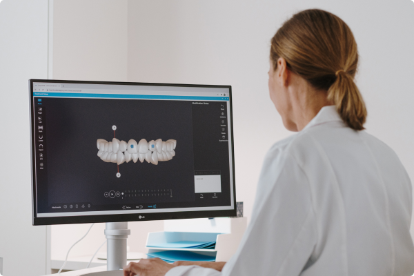
Patient communication
The ability to show the patient images of their oral anatomy both above and below the gums gives patients a much better understanding of the diagnosis and the intended treatment plan. Accurate pictures speak more than a thousand words which significantly improves patient understanding and therefore treatment uptake.Dental team communication
Digital images and CADCAM software have succeeded in improving communication between the whole dental team, including clinician, technician and any third-party partners. Communication is instant and misunderstandings are avoided because every team member has immediate access to the detailed images and design files. From a patient record-keeping point of view, digital images make it much easier to keep all patient information together in a single file on a practice management system for instance. Diagnostic images can then also be tracked more easily to monitor disease progression or any other changes in the patient’s dentition.